
What is it?
The canine stifle (knee) joint consists of an articulation between the femur (thigh bone) and tibia (shin bone). There are two cruciate ligaments within the stifle joint called the cranial and caudal cruciate ligaments which are fibrous bands that provide stability to the joint during weight bearing and twisting movements, and resisting movement between the femur and tibia during weight bearing. Cranial cruciate ligament disease is a common condition that affects dogs. This ligament may degenerate with age, weaken and then eventually rupture. Once stretched or ruptured (torn) the stifle becomes painful and unstable for the dog. Sometimes we see traumatic ruptures, but in most dogs the rupture occurs in a ligament with preexisting degenerative changes, often following minimal trauma.
We see complete or partial ruptures. Partial cruciate ruptures are unlikely to heal and may cause ongoing lameness or progress to complete ruptures. Degenerative changes may occur in the cruciate ligaments of both stifles, leading to simultaneous or successive rupture.
What are the clinical signs of cruciate disease in dogs?
Clinical signs of cruciate disease include hindlimb lameness, stifle swelling, pain and instability of the joint. Owners also often notice that dogs tend to sit ‘wonky’ or with the affected leg held out to the side. Thigh muscle loss may also occur.
In some cases, particularly where the meniscus is also injured (a shock absorber pad between the tibia and the femur), a clicking sound may be heard.
How is cranial cruciate ligament disease diagnosed?
Your dog will have an orthopaedic examination, which usually reveal signs of a joint effusion (an increase in the amount of fluid within the stifle), thickening of the joint and muscle wastage. Manipulation of the joint also allows the identification of instability. Following orthopaedic assessment, your dog will need to have a sedation or short general anaesthetic for examination of the stifle joint to further assess joint instability and for x-rays to be performed of the stifle joint, which can then be used to plan surgery.
Surgical Treatment
Surgical Treatment Surgical treatment for cruciate rupture is commonly recommended as it typically offers a superior outcome. At the time of surgery, the joint is inspected to assess the menisci and any injury is addressed. There are a number of surgical options each with individual advantages and disadvantages which will be discussed where appropriate.
Extra-capsular Repair: Lateral Fabellotibial Suture
This is a traditional treatment commonly employed, however it has in recent publications been demonstrated to offer not such a good outcome as TPLO (see Osteotomy Procedures) surgery particularly on medium to large breed dogs.
Complications are rare and the surgery is not as expensive as some of the other techniques. Surgery involves placement of a nylon implant on the outside of the joint to act as a temporary artificial cruciate ligament. The implant increases the stability of the stifle joint while tissue scarring occurs to further stabilise the joint. Failure of the implant before sufficient scarring is a common occurrence and may result in a prolonged or poor recovery.
Osteotomy Procedures
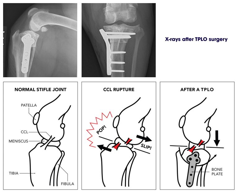
Osteotomy procedures typically involve cutting the shin bone to flatten the slope of the tibia (top of the shin), thereby removing the need to have a cruciate ligament to prevent movement of the femur (thigh bone) relative to the tibia. The cut bone is then stabilised with metal implants which hold the bone in the new position while healing occurs. The implants remain in place once healing has occurred unless there is a problem (e.g. infection necessitating their removal.
During the TPLO procedure a curved incision is made in the tibia which enables rotation and levelling of the tibial plateau. This is then secured in the new position using a bone plate and screws. The TPLO changes the shape of the joint so that the femur is less prone to slide down the tibial plateau when weight-bearing forces are applied; therefore improving the stability of the stifle joint. Recent publications have demonstrated that TPLO surgery offers the potential for a slightly superior outcome.
What could go wrong?
Potential complications will be discussed with you at consultation and include wound swelling or infection, surgical site infection, implant failure, fracture, ongoing lameness, late meniscal (cartilage joint pad) injury, patella tendon inflammation, osteoarthritis and ongoing lameness.
What is the likely outcome for my dog?
There is a good to excellent outcome for the majority of dogs following surgery after cruciate rupture. In a recent study, limb function of the operated leg of dogs receiving TPLO surgery was comparable in both walk and trot to the limb function of healthy dogs within 6-12 months of surgery. This was superior to the other techniques included in the study. Even though it is rarely clinically relevant, osteoarthritis keeps developing into the joint after surgery. Hence, for all dogs management relating to the treatment of osteoarthritis is advised. This includes dietary management, as obesity increases the risk of contralateral cruciate disease occurring, osteoarthritis progression and surgical complications.
Aftercare:
It is very important that you follow the post operative instructions carefully as failure to do so could result in the repair failing or the bone breaking, which potentially could result in further surgery being required and a prolonged recovery.
Exercise restriction:
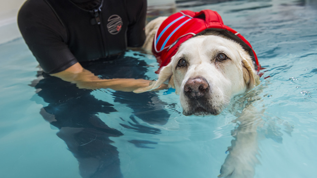
Unless instructed otherwise your dog should be rested in a crate, pen or small room for 6 to 8 weeks following surgery and should be walked on a short lead, four to five times daily for toileting only, then rested. Your dog should be consistently using the operated limb one week after surgery, and this should improve further over the coming weeks. Physiotherapy can be very beneficial for recovery following cruciate ligament surgery. Your pet may be discharged with a physiotherapy plan.
Follow up appointments
Unless advised otherwise, a revisit appointment with Langford Vets will be recommended in six to eight weeks for a check-up and postoperative x-rays to assess bone healing.
Returning your pet to exercise
Following your six week revisit and assuming your pet is making satisfactory progress, exercise can gradually be reintroduced. It is important to remember that the operated leg will still need to strengthen, and so failure to return to exercise gradually may result in complications. Hydrotherapy and / or physiotherapy can also be considered at this point if not already started.