
About Patellar Luxation
The patella (kneecap) runs in a groove on top of the femur, the movement of which is controlled by a tendon (fibrous band attached to the thigh muscles (quadriceps). The tendon and quadriceps muscles make up the quadriceps mechanism. Patellar luxation occurs when the patella moves out of the groove to sit on the inner or outer surface of the stifle (knee joint), called medial or lateral patellar luxation respectively.
Patellar luxation commonly occurs in small dogs, and also but less commonly large dogs. Patellar luxation is most commonly due to a developmental disorder caused by malalignment of the quadriceps mechanism and abnormalities of the thigh (femur) and shin (tibia) bones, with severe cases having a bowed appearance of the hindlimbs. Uncommonly patella luxation can be caused by a traumatic injury or following knee surgery. Patellar luxation is associated with the development of osteoarthritis in the stifle.
The severity of the patellar luxation is graded between 1 and 4:
• Grade 1
The patella can be pushed out of the groove, however immediately returns to a normal position, this isn’t generally associated with any clinical signs.
• Grade 2
The patella remains within the groove for the majority of the time but spontaneously moves out of the groove during limb movement.
• Grade 3
The patella is luxated out of the groove most of the time but can be replaced back into the groove.
• Grade 4
The patella is permanently luxated out of the groove and cannot be replaced.
Clinical Signs
Clinical signs of patellar luxation can be very variable. Common signs are an intermittent hopping lameness of the hindlimb during exercise, with one hindlimb held up for a few steps. Pets that are more severely affected may show a persistent continuous lameness.
How is Patellar Luxation Diagnosed?
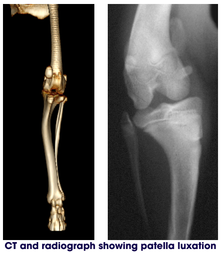
An orthopaedic examination of the conscious or sedated dog will be used to assess if patellar luxation is present and what grade it is. It will allow for assessment of any other orthopaedic conditions which are commonly present in animals with developmental patella luxation i.e. cranial cruciate ligament disease and hip dysplasia. Radiographs (x-rays) are taken to assess for concurrent problems, and for evidence of bone deformity. In more severe cases, a CT scan can be very useful to fully assess the deformity of the bones.
How is Patellar Luxation Diagnosed?
An orthopaedic examination of the conscious or sedated dog will be used to assess if patellar luxation is present and what grade it is. It will allow for assessment of any other orthopaedic conditions which are commonly present in animals with developmental patella luxation i.e. cranial cruciate ligament disease and hip dysplasia. Radiographs (x-rays) are taken to assess for concurrent problems, and for evidence of bone deformity. In more severe cases, a CT scan can be very useful to fully assess the deformity of the bones.
What are the treatment options?
Treatment will depend on the grade of luxation and the clinical signs.
Some dogs do not require surgery.
Conservative management can be tried which includes: weight management, exercise modification, physiotherapy, hydrotherapy and joint supplements. Conservative management will not stop patellar luxation but may assist in reducing the progression of associated osteoarthritis.
There are various surgical techniques, which are selected based on your pet’s individual anatomy, the direction of the patellar luxation, the severity of the lameness and whether there are any other hind limb conditions present.
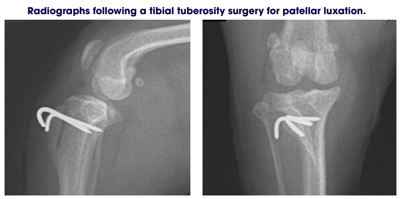
The aim of patellar luxation surgery is to stop the patella from luxating. Surgery is tailored to each case but typically involves improving the alignment of the quadriceps mechanism, by moving the tibial tuberosity (a small piece of bone at the top of the tibia) and deepening the groove the patella sits in. Then the soft tissues around the patella may be loosened, to allow the patella to sit back in the groove without tension, and overlapped to minimise the chance of reluxation.
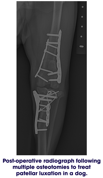
In more severe cases of patellar luxation it may be necessary to cut the femur to improve alignment of the bones. Metal implants are used to stabilise the cut bone whilst it heals.
What could go wrong?
Following surgery there is a low chance of complications. These include infection, swelling, reluxation of the patella, luxation of the patella in the other direction, breakage of the bones, implant loosening.
What is the likely outcome?
The prognosis following patellar luxation surgery is generally good. Osteoarthritis will occur in all dogs and cats to some degree, however this does not always cause lameness unless it becomes more severe. Some pets may benefit from ongoing conservative management including weight management, physiotherapy, hydrotherapy and joint supplements, which may slow the progression of osteoarthritis.
Exercise restriction
Following surgery it is essential for your pet to be rested whilst the bones and soft tissues heal. You will be given specific instructions tailored for your dog on your personalised discharge sheet. Generally for small-medium dogs we recommend crate rest. Larger dogs may be confined to a crate, small room, or a pen. If you do not already have a crate now is the time to start organising this. Exercise is usually restricted to short lead walks several times a day for a period of time.
Wound Care:
Following surgery the skin incision should be checked twice daily for two weeks or until the skin sutures have been removed. Licking or chewing of the surgical site can cause infection, and so we recommend that a buster collar be worn until the skin sutures are removed. If discharge or increased swelling is noted you should contact us and an examination may be necessary. Your dog may have been sent home with a small dressing in place over the incision. This can be left in place as long as it is clean and functional.
Physiotherapy and hydrotherapy:
Physiotherapy can be very beneficial for recovery following orthopaedic surgery. Where appropriate we will provide separate physiotherapy instructions.
Follow up appointments
Unless advised otherwise, a revisit appointment with Langford Vets will be recommended in six to eight weeks for a check-up and postoperative x-rays to assess bone healing.